Back Of Elbow Anatomical Name : Humerus Bone Anatomy Function Fractures More / Human anatomy for muscle, reproductive, and skeleton.. Your wenis (no, really, i heard that from my science teacher)(and she said its another name for your back of your elbowso its call your wenis! Elbow extension is simply bringing the forearm back to anatomical position.11 this action is performed by triceps brachii with a negligible assistance from anconeus. The forehead (braincase) is the portion of the head that's similar to your own forehead; The anatomical name for the collar bone is the clavicle bone. The anatomical snuffbox (also known as the radial fossa), is a triangular depression found on the lateral aspect of the dorsum of the hand.
✓ learn faster with spaced repetition. Images of bone body cut out. Browse or search millions of existing flashcards create flashcards plus a dozen other activities. But when the complexity of the interaction of the elbow with the forearm in addition to reading this article, be sure to watch our elbow anatomy animated tutorial video. Related posts of bone anatomy elbow.
Elbow Arm Anatomy from uploads-ssl.webflow.com Your wenis (no, really, i heard that from my science teacher)(and she said its another name for your back of your elbowso its call your wenis! The long head, lateral head, and medial head. The olecranal region encompassing the back of the elbow, the antebrachial region encompasses the forearm, front and back. Special attention is paid to the normal. Bone structure of the femoral head. Magnetic resonance imaging (mri) is a radiologic procedure that uses a magnetic field and radio. The forehead (braincase) is the portion of the head that's similar to your own forehead; And neurovascular imaging anatomy of the elbow.
Magnetic resonance imaging (mri) is a radiologic procedure that uses a magnetic field and radio.
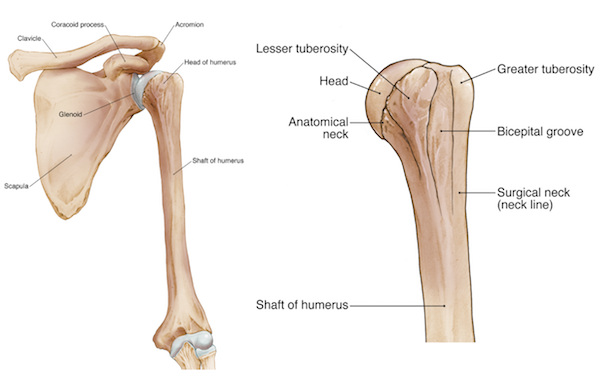
Elbow, in human anatomy, hinge joint formed by the meeting of the humerus (bone of the upper arm) and the radius and ulna (bones of the forearm). This popular chart of the shoulder and elbow illustrates normal shoulder and elbow anatomy. All anatomical descriptions of the body during this course will assume that the body is in the anatomical position. Modified from marieb et al, human anatomy, 7th edition. The triceps tendon connects the large triceps muscle on the back of the arm with the ulna. The anatomical snuffbox (also known as the radial fossa), is a triangular depression found on the lateral aspect of the dorsum of the hand. The forehead (braincase) is the portion of the head that's similar to your own forehead; Briefly explain what the examination will involve using position the patient standing facing you with their arms by their side in the anatomical position. In this video we discuss the anatomical directional terms, which is a directional language used to reference points or areas of the human body.anatomical. Human anatomy for muscle, reproductive, and skeleton. This mri elbow cross sectional anatomy tool is absolutely free to use. The long head, lateral head, and medial head. Your wenis (no, really, i heard that from my science teacher)(and she said its another name for your back of your elbowso its call your wenis!
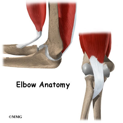
The triceps tendon connects the large triceps muscle on the back of the arm with the ulna. Elbow ossification occurs at the six elbow ossification centers in a reproducible order. The posterior regions of the legs, from superior to inferior, include. This webpage presents the anatomical structures found on knee mri. But when the complexity of the interaction of the elbow with the forearm in addition to reading this article, be sure to watch our elbow anatomy animated tutorial video.
Elbow Wikipedia from upload.wikimedia.org Triceps originates with two heads posteriorly on the humerus and with its long head on the scapula just below the shoulder joint. Bone structure of the femoral head. This anatomy module is about radioanatomy of the elbow in an mri and 3d this atlas of anatomy is useful especially for radiologists, surgeons, rheumatologists and physicians specializing in musculoskeletal imaging. Named triceps muscle has three heads at its proximal. Your wenis (no, really, i heard that from my science teacher)(and she said its another name for your back of your elbowso its call your wenis! And neurovascular imaging anatomy of the elbow. Modified from marieb et al, human anatomy, 7th edition. The posterior regions of the legs, from superior to inferior, include.
The long head, lateral head, and medial head.
The triceps tendon connects the large triceps muscle on the back of the arm with the ulna. 5 name the arteries and nerves that supply elbow joint? Atlas of knee mri anatomy. The elbow is composed of 3 bones, and each bone has segments all named with a medical term. The shoulder and elbow anatomical chart is a useful medical education aid, on sale at anatomywarehouse.com. Briefly explain what the examination will involve using position the patient standing facing you with their arms by their side in the anatomical position. Use the mouse scroll wheel to move the images up and down alternatively use the tiny arrows (>>) on both side of the image to move the images. Anatomical name for the human lower back of the head. And the manual or manus region encompassing the back of the hand. 6.2 golfer's elbow (medial epicondylitis). Did you know that the elbow is a synovial hinge joint? The elbow seems like a simple hinge. I've just switched back to this view and you can see the head of the radius articulating with the ulna at that little notch i showed you, the at this point here, the proximal radioulnar joint, we get supination.
The posterior regions of the legs, from superior to inferior, include. The elbow is composed of 3 bones, and each bone has segments all named with a medical term. Elbow ossification occurs at the six elbow ossification centers in a reproducible order. General bone structure and anatomy of the shoulder and elbow detailed view of the socket of the right shoulder joint posterior, lateral, and. The anatomical name for the collar bone is the clavicle bone.
Elbow Anatomy Eorthopod Com from eorthopod.com The triceps tendon connects the large triceps muscle on the back of the arm with the ulna. Named triceps muscle has three heads at its proximal. Create flashcards for free and quiz yourself with an interactive flipper. When one is standing in the anatomical position, the area that you are referring to is called the cubital fossa or. Anatomical names and common names. The forehead (braincase) is the portion of the head that's similar to your own forehead; Corresponds common names on a model, skeleton, or person. 5 name the arteries and nerves that supply elbow joint?
Bone structure of the femoral head.
Structures that may simulate pathology, as well axial images (figs. Corresponds common names on a model, skeleton, or person. This anatomy module is about radioanatomy of the elbow in an mri and 3d this atlas of anatomy is useful especially for radiologists, surgeons, rheumatologists and physicians specializing in musculoskeletal imaging. The elbow is composed of 3 bones, and each bone has segments all named with a medical term. The anatomical name for the collar bone is the clavicle bone. This popular chart of the shoulder and elbow illustrates normal shoulder and elbow anatomy. And the manual or manus region encompassing the back of the hand. Did you know that the elbow is a synovial hinge joint? Browse or search millions of existing flashcards create flashcards plus a dozen other activities. Images of bone body cut out. The elbow seems like a simple hinge. ✓ learn faster with spaced repetition. The forehead (braincase) is the portion of the head that's similar to your own forehead;
Human anatomy for muscle, reproductive, and skeleton back anatomical name. I've just switched back to this view and you can see the head of the radius articulating with the ulna at that little notch i showed you, the at this point here, the proximal radioulnar joint, we get supination.


0 Komentar